As a warning, some of the images in this post contain detailed depictions of human organs and anatomy.
Today, Collections Inventory Assistant, Immie unveils more secrets from the stores!
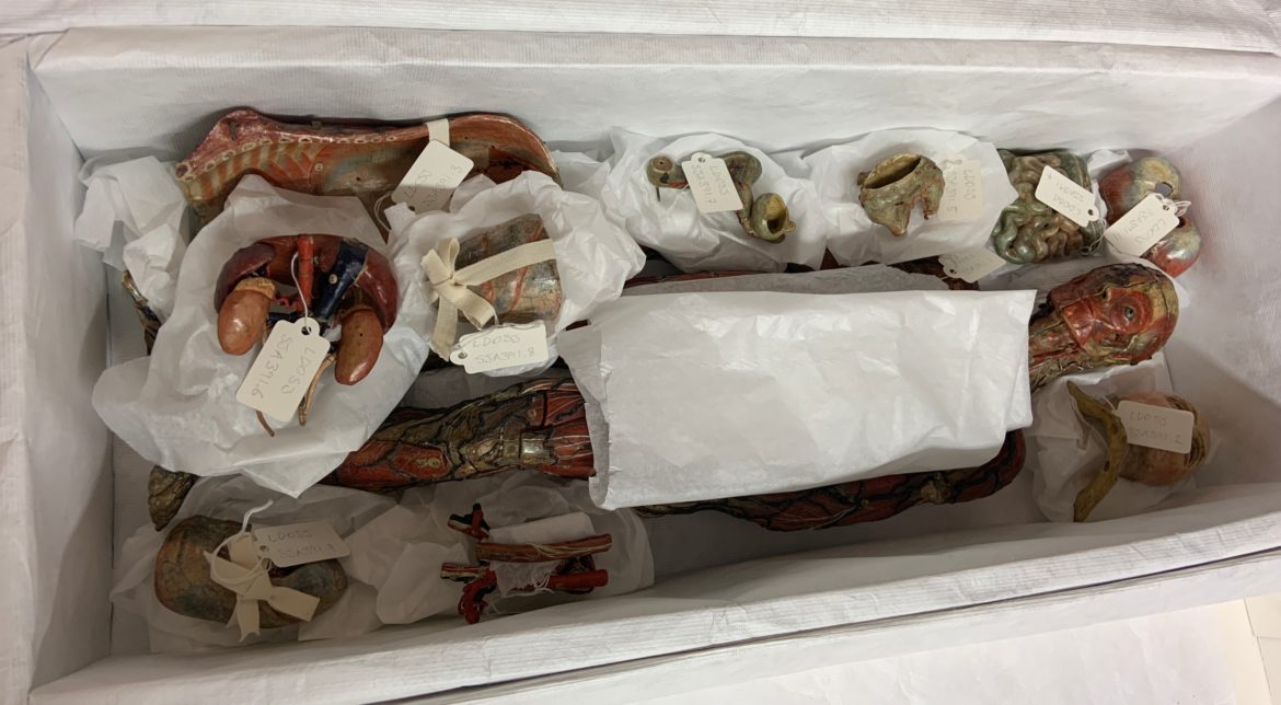
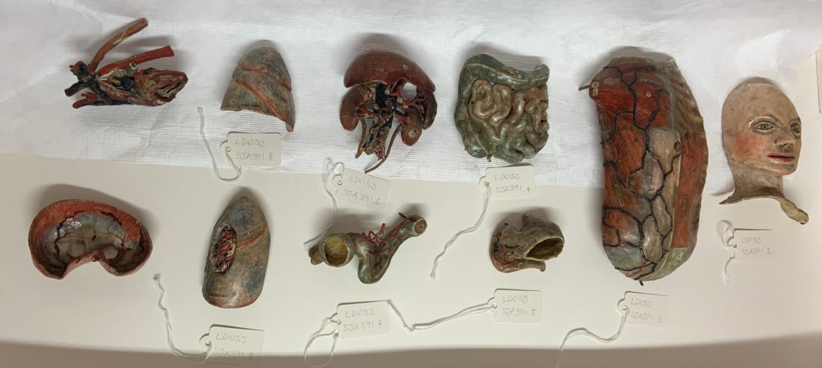
This object is an anatomical model of the human body and includes ten removeable organs, and even the face comes off! This object was donated to the museum by a member of the St John Ambulance Brigade. They were given the model by a doctor’s son whilst working in an air raid shelter in East London and used it for first aid training.


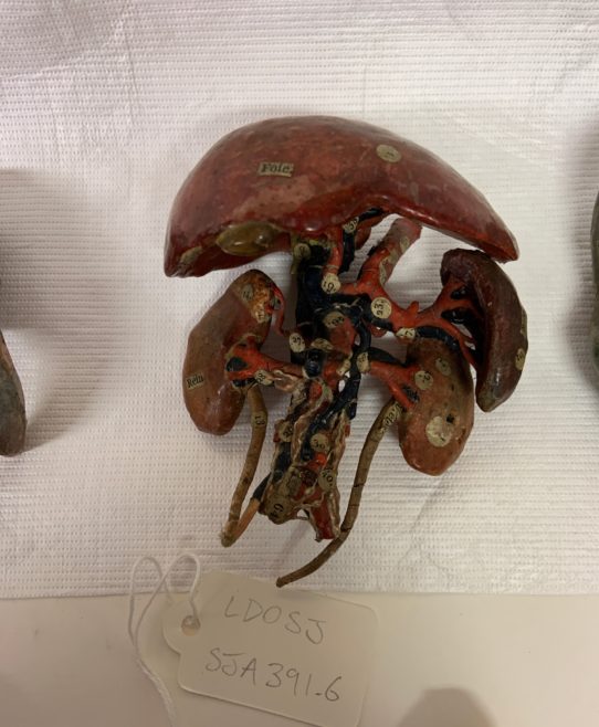
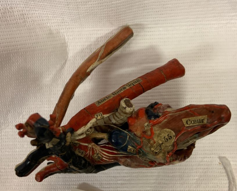
It is thought to have been made by the French physician and modelmaker, Louis Auzoux in the nineteenth century. These models were made from papier mâché, a new technique for the time, and highlighted veins, arteries and nerves with fabric covered wires. Anatomical models such as these were created in the nineteenth century in lieu of available corpses for dissection. In order to gain practical and working knowledge of the human anatomy, Auzoux created these models, often including removeable organs in order to mimic the process of dissection. These models became internationally renowned and feature in medical museum collections around the world.
The example from the Museum of the Order of St John’s collection is complete with small labels in French for each organ and body part. Each piece in this example is painted, varnished and labelled and includes small metal poles to fix the pieces in place. The high attention to detail and accuracy in this piece reveals the intent to use it as a teaching and learning model, where students could further their knowledge of the human body and its organs.

When this object was inventoried, it was decided that each piece should be given a separate number so we could record as much information and detail about the object as possible. To help preserve each part of the model, they were individually wrapped in acid-free tissue and labelled. This is to ensure they do not damage each other if the object is moved and any loose or delicate parts are properly cared for and the risk of damage is minimised.